Asanka Migelheva(@AMigelheva) 's Twitter Profile Photo

GNT Giant negative T ,defined as more than 10mm Deep T inversions (Yamaguchi et,al )

What are the important differentials when you see this rare ECG pattern ?

GNT Giant negative T ,defined as more than 10mm Deep T inversions (Yamaguchi et,al )

What are the important differentials when you see this rare ECG pattern ?
account_circle
Asanka Migelheva(@AMigelheva) 's Twitter Profile Photo

Easy case but still medical errors could happen
45 year female coming with acute onset central chest pain for the last 4 hours
Triage as CAT 2 , now machine concerned about acute ischaemia

Thoughts ?

Easy case but still medical errors could happen 
 45 year female coming with acute onset central chest pain for the last 4 hours 
Triage as CAT 2 , now machine concerned about acute ischaemia 

Thoughts ?
account_circle
Asanka Migelheva(@AMigelheva) 's Twitter Profile Photo

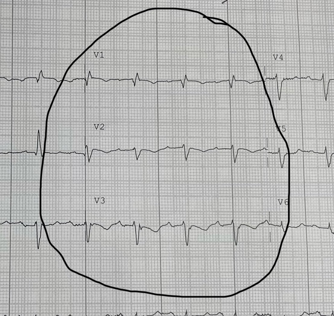
Patient with a syncope
S wave up stroke Will be more than 55 msec in V1 , V2

Which important cardiac pathology you would be concerned of ?

Patient with a syncope 
S wave up stroke Will be more than 55 msec in V1 , V2 

Which important cardiac pathology you would be concerned of ?
account_circle
Arron Pearce(@Arron_Pearce_) 's Twitter Profile Photo

Asanka Migelheva Screenshot below from Ken Grauer, MD's excellent blog, link: ecg-interpretation.blogspot.com/2013/01/ecg-in…

Takotsubo and CNS disorders are the only ones that tend to cause gross QT prolongation

@AMigelheva Screenshot below from @ekgpress's excellent blog, link: ecg-interpretation.blogspot.com/2013/01/ecg-in…

Takotsubo and CNS disorders are the only ones that tend to cause gross QT prolongation
account_circle
Asanka Migelheva(@AMigelheva) 's Twitter Profile Photo

81 year old BG of DM and Hypertension
4 hours Hx of acute central chest pain
ECG below .
She is otherwise stable.
Worried of an OMI ?

81 year old BG of DM and Hypertension 
4 hours Hx of acute central chest pain 
ECG below . 
She is otherwise stable. 
Worried of an OMI ?
account_circle
Asanka Migelheva(@AMigelheva) 's Twitter Profile Photo

Early repolarisation pattern in the ECG

Seems to be not so benign always
Matters in the Emergency department ?
Yes
Chest pain —- leads to diagnostic confusion

Syncope ——- think about ER syndrome

Early repolarisation pattern in the ECG 

Seems to be not so benign always 
Matters in the Emergency department ?
Yes 
Chest pain —- leads to diagnostic confusion 

Syncope ——- think about ER syndrome
account_circle
Dr Razi(@DrRazi4) 's Twitter Profile Photo

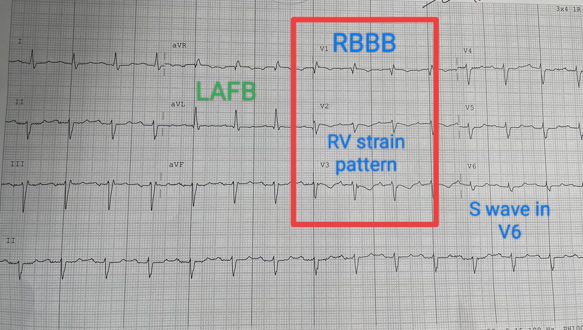
Features of RV enlargement;
1. Rbbb
2. RV strain pattern
3. S wave in V6

Bifascicular block - RBBB + LAFB

Possible diagnosis;
1. Acute PE
2. Chronic Respiratory Disease like OSA/OHS/COPD

#ECG Features of RV enlargement;
1. Rbbb 
2. RV strain pattern
3. S wave in V6

Bifascicular block - RBBB + LAFB

Possible diagnosis;
1. Acute PE 
2. Chronic Respiratory Disease like OSA/OHS/COPD
account_circle
Asanka Migelheva(@AMigelheva) 's Twitter Profile Photo

classical ECG changes which demarcate STEMI vs acute pericarditis “Pericarditis can cause localised ST elevation but there should be no reciprocal ST depression (except in AVR and V1)”

I have seen few cases which will challenge this teaching remotely
Your experiences ?

classical ECG changes which demarcate STEMI vs acute pericarditis “Pericarditis can cause localised ST elevation but there should be no reciprocal ST depression (except in AVR and V1)”

I have seen few cases which will challenge this teaching remotely 
Your experiences ?
account_circle
Asanka Migelheva(@AMigelheva) 's Twitter Profile Photo

24 M Hx of acute pericarditis 2 years back ( no records available )

Progressive exertion induced SOB over the past 6 months and now for past week having pleuritic type chest pain
What do you think about the ECG ?

24 M  Hx of acute pericarditis 2 years back ( no records available )

Progressive exertion induced SOB over the past 6 months and now for past week having pleuritic type chest pain 
What do you think about the ECG ?
account_circle
Dr Razi(@DrRazi4) 's Twitter Profile Photo

Asanka Migelheva Features of RV enlargement;
1. Rbbb
2. RV strain pattern
3. S wave in V6

Bifascicular block - RBBB + LAFB

Possible diagnosis;
1. Acute PE
2. Chronic Respiratory Disease like OSA/OHS/COPD

@AMigelheva Features of RV enlargement;
1. Rbbb 
2. RV strain pattern
3. S wave in V6

Bifascicular block - RBBB + LAFB

Possible diagnosis;
1. Acute PE 
2. Chronic Respiratory Disease like OSA/OHS/COPD
account_circle